
Advanced Bioinformatics and Biotechnology Approaches in Identifying and Targeting RNA Regulatory Networks
Bioinformatics in identifying and targeting RNA regulatory
DOI:
https://doi.org/10.66224/BiotechIntellect.3.1.64Keywords:
Non, coding RNA, RNA regulatory networks, Bioinformatics, Multi, omics integration, Experimental validation, RNA, targeting therapeuticsAbstract
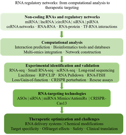
Background and Objective: RNA regulatory networks are of fundamental importance in gene expression, cellular homeostasis, development, and disease. Recent advances have improved the identification, characterization, and potential therapeutic targeting of these complex regulatory systems. This narrative review summarizes the computational and experimental approaches available for the study of RNA-mediated regulation with a particular focus on non-coding RNAs (ncRNAs), including miRNAs, lncRNAs, circRNAs, siRNAs, and piRNAs.
Materials and Methods: The review discusses computational tools and databases available for the prediction of RNA-RNA, RNA-protein, and transcription factor-RNA interactions, as well as the integration of multi-omics data to build regulatory networks. It goes on to consider the experimental technologies available for the profiling and validation of RNA interactions (bulk and single-cell RNA-seq, long-read sequencing, reporter assays, RIP, CLIP, and RNA-centric capture methods). Functional validation is also a major theme, with discussion of loss-of-function and gain-of-function experiments, CRISPR-based perturbation, rescue experiments, and the validation of feed-forward regulatory motifs. The review also considers the emerging biotechnologies for modulation of RNA regulatory networks (RNA-targeting technologies, CRISPR-based RNA modulation, delivery systems, and chemical modification of RNA therapeutics).
Results and Conclusion: By integrating computational prediction, multi-omics network construction, experimental validation, and emerging RNA-targeting biotechnologies, this review provides a comprehensive framework for translating RNA regulatory network discoveries into mechanistically supported and therapeutically relevant applications. Particular emphasis is placed on distinguishing predicted molecular associations from experimentally validated interactions and causal regulatory mechanisms.
Downloads
References
1. ENCODE Project Consortium, An integrated encyclopedia of DNA elements in the human genome. Nature, 2012. 489(7414): p. 57-74. https://doi.org/10.1038/nature11247
2. Statello, L., et al., Gene regulation by long non-coding RNAs and its biological functions. Nat Rev Mol Cell Biol, 2021. 22(2): p. 96-118. https://doi.org/10.1038/s41580-020-00315-9
3. Nemeth, K., et al., Non-coding RNAs in disease: from mechanisms to therapeutics. Nat Rev Genet, 2024. 25(3): p. 211-232. https://doi.org/10.1038/s41576-023-00662-1
4. Anastasiadou, E., L.S. Jacob, and F.J. Slack, Non-coding RNA networks in cancer. Nat Rev Cancer, 2018. 18(1): p. 5-18. https://doi.org/10.1038/nrc.2017.99
5. Yang, L.X., et al., The Application of Non-Coding RNAs as Biomarkers, Therapies, and Novel Vaccines in Diseases. Int J Mol Sci, 2025. 26(7): p. 3055. https://doi.org/10.3390/ijms26073055
6. Sato, K. and M. Hamada, Recent trends in RNA informatics: a review of machine learning and deep learning for RNA secondary structure prediction and RNA drug discovery. Brief Bioinform, 2023. 24(4): p. bbad186. https://doi.org/10.1093/bib/bbad186
7. Wang, K.C. and H.Y. Chang, Molecular mechanisms of long noncoding RNAs. Mol Cell, 2011. 43(6): p. 904-914. https://doi.org/10.1016/j.molcel.2011.08.018
8. Ding, S. and P. Wang, The Life of MicroRNAs: Biogenesis, Function and Decay in Cancer. Biomolecules, 2025. 15(10): p. 1393. https://doi.org/10.3390/biom15101393
9. Shang, R., et al., microRNAs in action: biogenesis, function and regulation. Nat Rev Genet, 2023. 24(12): p. 816-833. https://doi.org/10.1038/s41576-023-00611-y
10. Huntzinger, E. and E. Izaurralde, Gene silencing by microRNAs: contributions of translational repression and mRNA decay. Nat Rev Genet, 2011. 12(2): p. 99-110. https://doi.org/10.1038/nrg2936
11. O'Brien, J., et al., Overview of MicroRNA Biogenesis, Mechanisms of Actions, and Circulation. Front Endocrinol (Lausanne), 2018. 9: p. 402. https://doi.org/10.3389/fendo.2018.00402
12. Long, J. and F.R. Danesh, Promises and challenges of miRNA therapeutics. Am J Physiol Renal Physiol, 2022. 323(6): p. F673-f674. https://doi.org/10.1152/ajprenal.00251.2022
13. Sharma, S., A.A. Houfani, and L.J. Foster, Pivotal functions and impact of long non-coding RNAs on cellular processes and genome integrity. J Biomed Sci, 2024. 31(1): p. 52. https://doi.org/10.1186/s12929-024-01038-1
14. Rashid, F., A. Shah, and G. Shan, Long Non-coding RNAs in the Cytoplasm. Genomics Proteomics Bioinformatics, 2016. 14(2): p. 73-80. https://doi.org/10.1016/j.gpb.2016.03.005
15. Hwang, H.J. and Y.K. Kim, Molecular mechanisms of circular RNA translation. Exp Mol Med, 2024. 56(6): p. 1272-1280. https://doi.org/10.1038/s12276-024-01220-3
16. Asadi, M., et al., Therapeutic Potential of Circular RNAs as Targets for Cancer Treatment. Adv Pharm Bull, 2025. 15(3): p. 533-551. https://doi.org/10.34172/apb.025.45330
17. Margvelani, G., et al., Translation of circular RNAs. Nucleic Acids Res, 2025. 53(1): p. gkae1167. https://doi.org/10.1093/nar/gkae1167
18. Idda, M.L., A.F. Fara, and M. Floris, Circular RNAs in Immune-Related Diseases. Adv Exp Med Biol, 2025. 1485: p. 417-435. https://doi.org/10.1007/978-981-96-9428-0_24
19. Wilson, R.C. and J.A. Doudna, Molecular mechanisms of RNA interference. Annu Rev Biophys, 2013. 42: p. 217-239. https://doi.org/10.1146/annurev-biophys-083012-130404
20. Dana, H., et al., Molecular Mechanisms and Biological Functions of siRNA. Int J Biomed Sci, 2017. 13(2): p. 48-57. https://doi.org/10.59566/ijbs.2017.13048
21. Hu, B., et al., Therapeutic siRNA: state of the art. Signal Transduct Target Ther, 2020. 5(1): p. 101. https://doi.org/10.1038/s41392-020-0207-x
22. Fu, Q. and P.J. Wang, Mammalian piRNAs: Biogenesis, function, and mysteries. Sperma-togenesis, 2014. 4: p. e27889. https://doi.org/10.4161/spmg.27889
23. Weick, E.M. and E.A. Miska, piRNAs: from biogenesis to function. Development, 2014. 141(18): p. 3458-3471. https://doi.org/10.1242/dev.094037
24. Winkle, M., et al., Noncoding RNA therapeutics - challenges and potential solutions. Nat Rev Drug Discov, 2021. 20(8): p. 629-651. https://doi.org/10.1038/s41573-021-00219-z
25. Rincón-Riveros, A., et al., Bioinformatic Tools for the Analysis and Prediction of ncRNA Interactions. Int J Mol Sci, 2021. 22(21): p. 11397. https://doi.org/10.3390/ijms222111397
26. Teng, Y., et al., Associative Analysis of lncRNA/circRNA-miRNA-mRNA Expression Pro-files in Iron-Overloaded HT-1080 Fibrosarcoma Cells. Int J Mol Sci, 2026. 27(12): p. 5617. https://doi.org/10.3390/ijms27125617
27. Ragusa, M., et al., Molecular Crosstalking among Noncoding RNAs: A New Network Layer of Genome Regulation in Cancer. Int J Genomics, 2017. 2017: p. 4723193. https://doi.org/10.1155/2017/4723193
28. Singh, A., et al., Advancements in RNA-based therapies from bench to bedside. NPJ Drug Discov, 2026. 3(1): p. 4. https://doi.org/10.1038/s44386-025-00037-y
29. Skaineh, Z., et al., A Narrative Review on Integrative Bioinformatics Approaches for microRNA Research in Familial Mediterranean Fever: Current Insights and Future Directions. Health Sci Rep, 2026. 9(3): p. e72047. https://doi.org/10.1002/hsr2.72047
30. Liu, B., J. Li, and M.J. Cairns, Identifying miRNAs, targets and functions. Brief Bioinform, 2014. 15(1): p. 1-19. https://doi.org/10.1093/bib/bbs075
31. Hall, M.H., et al., Functional microRNA targeting without seed pairing. Nucleic Acids Res, 2025. 53(19): p. gkaf1018. https://doi.org/10.1093/nar/gkaf1018
32. Song, Y., et al., ceRNAshiny: An Interactive R/Shiny App for Identification and Analysis of ceRNA Regulation. Front Mol Biosci, 2022. 9: p. 865408. https://doi.org/10.3389/fmolb.2022.865408
33. Bang, D., et al., A Survey on Computational Methods for Investigation on ncRNA-Disease Association through the Mode of Action Perspective. Int J Mol Sci, 2022. 23(19): p. 11498. https://doi.org/10.3390/ijms231911498
34. Riolo, G., et al., miRNA Targets: From Prediction Tools to Experimental Validation. Methods Protoc, 2020. 4(1): p. 1. https://doi.org/10.3390/mps4010001
35. Betel, D., et al., Comprehensive modeling of microRNA targets predicts functional non-conserved and non-canonical sites. Genome Biol, 2010. 11(8): p. R90. https://doi.org/10.1186/gb-2010-11-8-r90
36. Shakyawar, S., S. Southekal, and C. Guda, mintRULS: Prediction of miRNA-mRNA Target Site Interactions Using Regularized Least Square Method. Genes (Basel), 2022. 13(9): p. 1528. https://doi.org/10.3390/genes13091528
37. Pinzón, N., et al., microRNA target prediction programs predict many false positives. Genome Res, 2017. 27(2): p. 234-245. https://doi.org/10.1101/gr.205146.116
38. Vejnar, C.E. and E.M. Zdobnov, miRmap: Comprehensive prediction of microRNA target repression strength. Nucleic Acids Res, 2012. 40(22): p. 11673-11683. https://doi.org/10.1093/nar/gks901
39. Chen, Y. and X. Wang, miRDB: an online database for prediction of functional microRNA targets. Nucleic Acids Res, 2020. 48(D1): p. D127-d131. https://doi.org/10.1093/nar/gkz757
40. Wong, N. and X. Wang, miRDB: an online resource for microRNA target prediction and functional annotations. Nucleic Acids Res, 2015. 43(Database issue): p. D146-152. https://doi.org/10.1093/nar/gku1104
41. Clark, A.J. and J.W. Lillard, Jr., A Comprehensive Review of Bioinformatics Tools for Genomic Biomarker Discovery Driving Precision Oncology. Genes (Basel), 2024. 15(8). https://doi.org/10.3390/genes15081036
42. Chang, L., et al., miRNet 2.0: network-based visual analytics for miRNA functional analysis and systems biology. Nucleic Acids Res, 2020. 48(W1): p. W244-w251. https://doi.org/10.1093/nar/gkaa467
43. Tastsoglou, S., et al., DIANA-microT 2023: including predicted targets of virally encoded miRNAs. Nucleic Acids Res, 2023. 51(W1): p. W148-w153. https://doi.org/10.1093/nar/gkad283
44. Skoufos, G., et al., TarBase-v9.0 extends experimentally supported miRNA–gene interactions to cell-types and virally encoded miRNAs. Nucleic Acids Res, 2024. 52(D1): p. D304-D310. https://doi.org/10.1093/nar/gkad1071
45. Gao, Y., et al., Recent advances in investigation of circRNA/lncRNA-miRNA-mRNA networks through RNA sequencing data analysis. Brief Funct Genomics, 2025. 24: p. elaf005. https://doi.org/10.1093/bfgp/elaf005
46. Paraskevopoulou, M.D., et al., DIANA-LncBase: experimentally verified and computationally predicted microRNA targets on long non-coding RNAs. Nucleic Acids Res, 2013. 41(Database issue): p. D239-245. https://doi.org/10.1093/nar/gks1246
47. Li, J.H., et al., starBase v2.0: decoding miRNA-ceRNA, miRNA-ncRNA and protein-RNA interaction networks from large-scale CLIP-Seq data. Nucleic Acids Res, 2014. 42(Database issue): p. D92-97. https://doi.org/10.1093/nar/gkt1248
48. Kang, J., et al., RNAInter v4.0: RNA interactome repository with redefined confidence scoring system and improved accessibility. Nucleic Acids Res, 2022. 50(D1): p. D326-d332. https://doi.org/10.1093/nar/gkab997
49. Wang, J., et al., TransmiR: a transcription factor-microRNA regulation database. Nucleic Acids Res, 2010. 38(Database issue): p. D119-122. https://doi.org/10.1093/nar/gkp803
50. Li, Y., et al., Systematic review of computational methods for identifying miRNA-mediated RNA-RNA crosstalk. Brief Bioinform, 2019. 20(4): p. 1193-1204. https://doi.org/10.1093/bib/bbx137
51. Fabbri, M., et al., Decrypting noncoding RNA interactions, structures, and functional networks. Genome Res, 2019. 29(9): p. 1377-1388. https://doi.org/10.1101/gr.247239.118
52. Mann, M., P.R. Wright, and R. Backofen, IntaRNA 2.0: enhanced and customizable prediction of RNA-RNA interactions. Nucleic Acids Res, 2017. 45(W1): p. W435-w439. https://doi.org/10.1093/nar/gkx279
53. Aborode, A.T., et al., RNA binding proteins (RBPs) on genetic stability and diseases. Glob Med Genet, 2025. 12(1): p. 100032. https://doi.org/10.1016/j.gmg.2024.100032
54. De, S. and M. Gorospe, Bioinformatic tools for analysis of CLIP ribonucleoprotein data. Wiley Interdiscip Rev RNA, 2017. 8(4). https://doi.org/10.1002/wrna.1404
55. Lin, Y., et al., RNAInter in 2020: RNA interactome repository with increased coverage and annotation. Nucleic Acids Res, 2020. 48(D1): p. D189-D197. https://doi.org/10.1093/nar/gkz804
56. Lu, Q., et al., Identification of potentially functional circRNAs and prediction of the circRNA-miRNA-hub gene network in mice with primary blast lung injury. BMC Pulm Med, 2023. 23(1): p. 410. https://doi.org/10.1186/s12890-023-02717-9
57. Hoffmann, M., et al., circRNA-sponging: a pipeline for extensive analysis of circRNA expression and their role in miRNA sponging. Bioinform Adv, 2023. 3(1): p. vbad093. https://doi.org/10.1093/bioadv/vbad093
58. Tong, Y., J.L. Childs‐Disney, and M.D. Disney, Targeting RNA with small molecules, from RNA structures to precision medicines: IUPHAR review: 40. Br J Pharmacol, 2024. 181(21): p. 4152-4173. https://doi.org/10.1111/bph.17308
59. Budnik, M., et al., Deep dive into RNA: a systematic literature review on RNA structure prediction using machine learning methods: M. Budnik et al. Artif Intell Rev, 2024. 57(9): p. 254. https://doi.org/10.1007/s10462-024-10910-3
60. Wang, S., D. Weissman, and Y. Dong, RNA chemistry and therapeutics. Nat Rev Drug Discov, 2025. 24(11): p. 828-851. https://doi.org/10.1038/s41573-025-01237-x
61. Tang, Q. and A. Khvorova, RNAi-based drug design: considerations and future directions. Nat Rev Drug Discov, 2024. 23(5): p. 341-364. https://doi.org/10.1038/s41573-024-00912-9
62. Jiang, W., et al., Network-based multi-omics integrative analysis methods in drug discovery: a systematic review. BioData Min, 2025. 18(1): p. 27. https://doi.org/10.1186/s13040-025-00442-z
63. Monfort-Lanzas, P., et al., Machine learning to dissect perturbations in complex cellular systems. 2025: p. 832-842. https://doi.org/10.1016/j.csbj.2025.02.028
64. Conesa, A., et al., A survey of best practices for RNA-seq data analysis. Genome Biol, 2016. 17(1): p. 13. https://doi.org/10.1186/s13059-016-0881-8
65. Kalisky, T., et al., A brief review of single-cell transcriptomic technologies. Brief Funct Genomics, 2018. 17(1): p. 64-76. https://doi.org/10.1093/bfgp/elx019
66. Markowetz, F. and R. Spang, Inferring cellular networks--a review. BMC Bioinformatics, 2007. 8 Suppl 6(Suppl 6): p. S5. https://doi.org/10.1186/1471-2105-8-s6-s5
67. Wang, Z., M. Gerstein, and M. Snyder, RNA-Seq: a revolutionary tool for transcriptomics. Nat Rev Genet, 2009. 10(1): p. 57-63. https://doi.org/10.1038/nrg2484
68. Langfelder, P. and S. Horvath, WGCNA: an R package for weighted correlation network analysis. BMC Bioinformatics, 2008. 9: p. 559. https://doi.org/10.1186/1471-2105-9-559
69. Kuhn, A., et al., Population-specific expression analysis (PSEA) reveals molecular changes in diseased brain. Nat Methods, 2011. 8(11): p. 945-947. https://doi.org/10.1038/nmeth.1710
70. Baran-Gale, J., et al., Addressing Bias in Small RNA Library Preparation for Sequencing: A New Protocol Recovers MicroRNAs that Evade Capture by Current Methods. Front Genet, 2015. 6: p. 352. https://doi.org/10.3389/fgene.2015.00352
71. Benesova, S., M. Kubista, and L. Valihrach, Small RNA-Sequencing: Approaches and Considerations for miRNA Analysis. Diagnostics (Basel), 2021. 11(6): p. 964. https://doi.org/10.3390/diagnostics11060964
72. Giraldez, M.D., et al., Comprehensive multi-center assessment of small RNA-seq methods for quantitative miRNA profiling. Nat Biotechnol, 2018. 36(8): p. 746-757. https://doi.org/10.1038/nbt.4183
73. Saliba, A.E., et al., Single-cell RNA-seq: advances and future challenges. Nucleic Acids Res, 2014. 42(14): p. 8845-8860. https://doi.org/10.1093/nar/gku555
74. Potter, S.S., Single-cell RNA sequencing for the study of development, physiology and disease. Nat Rev Nephrol, 2018. 14(8): p. 479-492. https://doi.org/10.1038/s41581-018-0021-7
75. Haque, A., et al., A practical guide to single-cell RNA-sequencing for biomedical research and clinical applications. Genome Med, 2017. 9(1): p. 75. https://doi.org/10.1186/s13073-017-0467-4
76. Ament, I.H., et al., Long-read RNA sequencing: A transformative technology for exploring transcriptome complexity in human diseases. Mol Ther, 2025. 33(3): p. 883-894. https://doi.org/10.1016/j.ymthe.2024.11.025
77. Lillebæk, E.M.S. and B.H. Kallipolitis, Experimental Validation of RNA-RNA Interactions by Electrophoretic Mobility Shift Assay. Methods Mol Biol, 2024. 2741: p. 195-206. https://doi.org/10.1007/978-1-0716-3565-0_11
78. Song, Z. and E.L. Van Nostrand, Progress and challenges in profiling protein-RNA and protein-associated RNA-RNA interactions. RNA, 2026. 32(4): p. 457-471. https://doi.org/10.1261/rna.080830.125
79. Clément, T., V. Salone, and M. Rederstorff, Dual luciferase gene reporter assays to study miRNA function. Methods Mol Biol, 2015. 1296: p. 187-198. https://doi.org/10.1007/978-1-4939-2547-6_17
80. Nechay, M. and R.E. Kleiner, High-throughput approaches to profile RNA-protein interactions. Curr Opin Chem Biol, 2020. 54: p. 37-44. https://doi.org/10.1016/j.cbpa.2019.11.002
81. Helwak, A., et al., Mapping the human miRNA interactome by CLASH reveals frequent noncanonical binding. Cell, 2013. 153(3): p. 654-665. https://doi.org/10.1016/j.cell.2013.03.043
82. Moore, M.J., et al., miRNA-target chimeras reveal miRNA 3'-end pairing as a major determinant of Argonaute target specificity. Nat Commun, 2015. 6: p. 8864. https://doi.org/10.1038/ncomms9864
83. Cabili, M.N., et al., Localization and abundance analysis of human lncRNAs at single-cell and single-molecule resolution. Genome Biol, 2015. 16(1): p. 20. https://doi.org/10.1186/s13059-015-0586-4
84. Eliscovich, C., S.M. Shenoy, and R.H. Singer, Imaging mRNA and protein interactions within neurons. Proc Natl Acad Sci U S A, 2017. 114(10): p. E1875-e1884. https://doi.org/10.1073/pnas.1621440114
85. Barra, J., R. Vendramin, and E. Leucci, M(R)apping RNA-Protein Interactions. Methods Mol Biol, 2021. 2254: p. 259-272. https://doi.org/10.1007/978-1-0716-1158-6_16
86. Gagliardi, M. and M.R. Matarazzo, RIP: RNA Immunoprecipitation. Methods Mol Biol, 2016. 1480: p. 73-86. https://doi.org/10.1007/978-1-4939-6380-5_7
87. Ule, J., et al., CLIP identifies Nova-regulated RNA networks in the brain. Science, 2003. 302(5648): p. 1212-1215. https://doi.org/10.1126/science.1090095
88. Van Nostrand, E.L., et al., Robust transcriptome-wide discovery of RNA-binding protein binding sites with enhanced CLIP (eCLIP). Nat Methods, 2016. 13(6): p. 508-514. https://doi.org/10.1038/nmeth.3810
89. Ramanathan, M., D.F. Porter, and P.A. Khavari, Methods to study RNA-protein interactions. Nat Methods, 2019. 16(3): p. 225-234. https://doi.org/10.1038/s41592-019-0330-1
90. Mattick, J.S., et al., Long non-coding RNAs: definitions, functions, challenges and recommendations. Nat Rev Mol Cell Biol, 2023. 24(6): p. 430-447. https://doi.org/10.1038/s41580-022-00566-8
91. Pamudurti, N.R., et al., Translation of CircRNAs. Mol Cell, 2017. 66(1): p. 9-21.e7. https://doi.org/10.1016/j.molcel.2017.02.021
92. Bhardwaj, A.R., et al., Northern blotting technique for detection and expression analysis of mRNAs and small RNAs, in RNA Abundance Analysis: Methods and Protocols. 2020, Springer. p. 155-183: p. 155-183. https://doi.org/10.1007/978-1-0716-0743-5_12
93. Memczak, S., et al., Circular RNAs are a large class of animal RNAs with regulatory potency. Nature, 2013. 495(7441): p. 333-338. https://doi.org/10.1038/nature11928
94. Nielsen, A.F., et al., Best practice standards for circular RNA research. Nat Methods, 2022. 19(10): p. 1208-1220. https://doi.org/10.1038/s41592-022-01487-2
95. Raj, A., et al., Imaging individual mRNA molecules using multiple singly labeled probes. Nat Methods, 2008. 5(10): p. 877-879. https://doi.org/10.1038/nmeth.1253
96. Sang, L., et al., Subcellular distribution, localization, and function of noncoding RNAs. Wiley Interdiscip Rev RNA, 2022. 13(6): p. e1729. https://doi.org/10.1002/wrna.1729
97. Zubradt, M., et al., DMS-MaPseq for genome-wide or targeted RNA structure probing in vivo. Nat Methods, 2017. 14(1): p. 75-82. https://doi.org/10.1038/nmeth.4057
98. Spitale, R.C. and D. Incarnato, Probing the dynamic RNA structurome and its functions. Nat Rev Genet, 2023. 24(3): p. 178-196. https://doi.org/10.1038/s41576-022-00546-w
99. Guttman, M., et al., Chromatin signature reveals over a thousand highly conserved large non-coding RNAs in mammals. Nature, 2009. 458(7235): p. 223-227. https://doi.org/10.1038/nature07672
100. Tsai, M.C., et al., Long noncoding RNA as modular scaffold of histone modification complexes. Science, 2010. 329(5992): p. 689-693. https://doi.org/10.1126/science.1192002
101. Kato, M. and P. Carninci, Genome-Wide Technologies to Study RNA-Chromatin Inter-actions. Noncoding RNA, 2020. 6(2): p. 20. https://doi.org/10.3390/ncrna6020020
102. Park, P.J., ChIP-seq: advantages and challenges of a maturing technology. Nat Rev Genet, 2009. 10(10): p. 669-680. https://doi.org/10.1038/nrg2641
103. Marson, A., et al., Connecting microRNA genes to the core transcriptional regulatory circuitry of embryonic stem cells. Cell, 2008. 134(3): p. 521-533. https://doi.org/10.1016/j.cell.2008.07.020
104. Furey, T.S., ChIP-seq and beyond: new and improved methodologies to detect and characterize protein-DNA interactions. Nat Rev Genet, 2012. 13(12): p. 840-852. https://doi.org/10.1038/nrg3306
105. Chu, C., J. Quinn, and H.Y. Chang, Chromatin isolation by RNA purification (ChIRP). J Vis Exp, 2012(61). https://doi.org/10.3791/3912
106. Cao, M., J. Zhao, and G. Hu, Genome-wide methods for investigating long noncoding RNAs. Biomed Pharmacother, 2019. 111: p. 395-401. https://doi.org/10.1016/j.biopha.2018.12.078
107. Simon, M.D., et al., High-resolution Xist binding maps reveal two-step spreading during X-chromosome inactivation. Nature, 2013. 504(7480): p. 465-469. https://doi.org/10.1038/nature12719
108. Engreitz, J.M., et al., The Xist lncRNA exploits three-dimensional genome architecture to spread across the X chromosome. Science, 2013. 341(6147): p. 1237973. https://doi.org/10.1126/science.1237973
109. Engreitz, J., E.S. Lander, and M. Guttman, RNA antisense purification (RAP) for mapping RNA interactions with chromatin, in Nuclear Bodies and Noncoding RNAs: Methods and Protocols. 2014, Springer. p. 183-197: p. 183-197. https://doi.org/10.1007/978-1-4939-2253-6_11
110. Kopp, F. and J.T. Mendell, Functional Classif-ication and Experimental Dissection of Long Noncoding RNAs. Cell, 2018. 172(3): p. 393-407. https://doi.org/10.1016/j.cell.2018.01.011
111. Konermann, S., et al., Genome-scale trans-criptional activation by an engineered CRISPR-Cas9 complex. Nature, 2015. 517(7536): p. 583-588. https://doi.org/10.1038/nature14136
112. Abudayyeh, O.O., et al., RNA targeting with CRISPR-Cas13. Nature, 2017. 550(7675): p. 280-284. https://doi.org/10.1038/nature24049
113. Cox, D.B.T., et al., RNA editing with CRISPR-Cas13. Science, 2017. 358(6366): p. 1019-1027. https://doi.org/10.1126/science.aaq0180
114. Goyal, A., et al., Challenges of CRISPR/Cas9 applications for long non-coding RNA genes. Nucleic Acids Res, 2017. 45(3): p. e12. https://doi.org/10.1093/nar/gkw883
115. Tsang, J., J. Zhu, and A. van Oudenaarden, MicroRNA-mediated feedback and feedforward loops are recurrent network motifs in mammals. Mol Cell, 2007. 26(5): p. 753-767. https://doi.org/10.1016/j.molcel.2007.05.018
116. Saviano, A., N.C. Henderson, and T.F. Baumert, Single-cell genomics and spatial transcriptomics: Discovery of novel cell states and cellular interactions in liver physiology and disease biology. J Hepatol, 2020. 73(5): p. 1219-1230. https://doi.org/10.1016/j.jhep.2020.06.004
117. Kuhn, D.E., et al., Experimental validation of miRNA targets. Methods, 2008. 44(1): p. 47-54. https://doi.org/10.1016/j.ymeth.2007.09.005
118. Van Nostrand, E.L., et al., A large-scale binding and functional map of human RNA-binding proteins. Nature, 2020. 583(7818): p. 711-719. https://doi.org/10.1038/s41586-020-2077-3
119. McHugh, C.A., P. Russell, and M. Guttman, Methods for comprehensive experimental identification of RNA-protein interactions. Genome Biol, 2014. 15(1): p. 203. https://doi.org/10.1186/gb4152
120. Crooke, S.T., et al., Antisense technology: an overview and prospectus. Nat Rev Drug Discov, 2021. 20(6): p. 427-453. https://doi.org/10.1038/s41573-021-00162-z
121. Jadhav, V., et al., RNA interference in the era of nucleic acid therapeutics. Nat Biotechnol, 2024. 42(3): p. 394-405. https://doi.org/10.1038/s41587-023-02105-y
122. Rupaimoole, R. and F.J. Slack, MicroRNA therapeutics: towards a new era for the management of cancer and other diseases. Nat Rev Drug Discov, 2017. 16(3): p. 203-222. https://doi.org/10.1038/nrd.2016.246
123. Yang, H. and D.J. Patel, Structures, mechanisms and applications of RNA-centric CRISPR–Cas13. Nat Chem Biol, 2024. 20(6): p. 673-688. https://doi.org/10.1038/s41589-024-01593-6
124. Konermann, S., et al., Transcriptome engineering with RNA-targeting type VI-D CRISPR effectors. Cell, 2018. 173(3): p. 665-676.e14. https://doi.org/10.1016/j.cell.2018.02.033
125. Liang, W.-W., et al., Transcriptome-scale RNA-targeting CRISPR screens reveal essential lncRNAs in human cells. Cell, 2024. 187(26): p. 7637-7654.e29. https://doi.org/10.1016/j.cell.2024.10.021
126. Shi, P. and X. Wu, Programmable RNA targeting with CRISPR-Cas13. RNA Biol, 2024. 21(1): p. 1-9. https://doi.org/10.1080/15476286.2024.2351657
127. Moazzam, M., et al., The landscape of nanoparticle-based siRNA delivery and therapeutic development. Mol Ther, 2024. 32(2): p. 284-312. https://doi.org/10.1016/j.ymthe.2024.01.005
128. Hou, X., et al., Lipid nanoparticles for mRNA delivery. Nat Rev Mater, 2021. 6(12): p. 1078-1094. https://doi.org/10.1038/s41578-021-00358-0
129. Song, D., et al., Tuning Lipid Nanoparticles for RNA Delivery to Extrahepatic Organs. Adv Mater, 2024. 36(44): p. e2401445. https://doi.org/10.1002/adma.202401445
130. Paunovska, K., D. Loughrey, and J.E. Dahlman, Drug delivery systems for RNA therapeutics. Nat Rev Genet, 2022. 23(5): p. 265-280. https://doi.org/10.1038/s41576-021-00439-4
131. Juliano, R.L., Intracellular trafficking and endosomal release of oligonucleotides: what we know and what we don’t. Nucleic Acid Ther, 2018. 28(3): p. 166-177. https://doi.org/10.1089/nat.2018.0727
132. Shen, X. and D.R. Corey, Chemistry, mechanism and clinical status of antisense oligonucleotides and duplex RNAs. Nucleic Acids Res, 2018. 46(4): p. 1584-1600. https://doi.org/10.1093/nar/gkx1239
133. Shadid, M., M. Badawi, and A. Abulrob, Antisense oligonucleotides: absorption, distribution, metabolism, and excretion. Expert Opin Drug Metab Toxicol, 2021. 17(11): p. 1281-1292. https://doi.org/10.1080/17425255.2021.1992382
134. Crooke, S.T., et al., Antisense technology: A review. J Biol Chem, 2021. 296: p. 100416. https://doi.org/10.1016/j.jbc.2021.100416
135. Burel, S.A., et al., Hepatotoxicity of high affinity gapmer antisense oligonucleotides is mediated by RNase H1 dependent promiscuous reduction of very long pre-mRNA transcripts. Nucleic Acids Res, 2016. 44(5): p. 2093-2109. https://doi.org/10.1093/nar/gkv1210
136. Egli, M. and M. Manoharan, Chemistry, structure and function of approved oligonucleotide therapeutics. Nucleic Acids Res, 2023. 51(6): p. 2529-2573. https://doi.org/10.1093/nar/gkad067
137. Csermely, P., et al., Structure and dynamics of molecular networks: a novel paradigm of drug discovery: a comprehensive review. Pharmacol Ther, 2013. 138(3): p. 333-408. https://doi.org/10.1016/j.pharmthera.2013.01.016
138. Lee, D. and S. Kim, Knowledge-guided artificial intelligence technologies for decoding complex multiomics interactions in cells. Clin Exp Pediatr, 2022. 65(5): p. 239-249. https://doi.org/10.3345/cep.2021.01438
Downloads
Publication Timeline
- Submitted
- Revised
- Accepted
Issue
Section
License
Copyright (c) 2026 Nasrin Amiri-Dashatan, Mahdis Nazari , Mehdi Koushki , Lobat Tayebi (Author); Masoumeh Farahani, Mostafa Rezaei-Tavirani

This work is licensed under a Creative Commons Attribution-NonCommercial 4.0 International License.